A paper describing our collaborative work with Dr Joel Boerckel’s group in the University of Pennsylvania, USA has been published in Developmental Dynamics! Congratulations to lead author Dr Christopher Panebianco and all co-authors!
In the research, we investigate how the bones of the lower forelimb of the goat develop from after birth until maturity (older than two years for the goat).
What we found was that the cortical (shell) of the first phalangeal bone (P1) bone – the first digit in the forefoot of the goat- increases in density quickly after birth, while the thickness of that shell increases continually until skeletal maturity. The polar moment of inertia of the bone (a measure of how easy or hard the bone is to twist) is constant over time, indicating that the mechanical performance of the bone adapts along with the weight of the goat as it grows. In the trabecular (spongy) bone, we found that the number and thickness of the trabeculae increase until sexual maturity (12 months) in the P1 bone, while the trabeculae in the metacarpal bone increase in thickness and not as much in number.
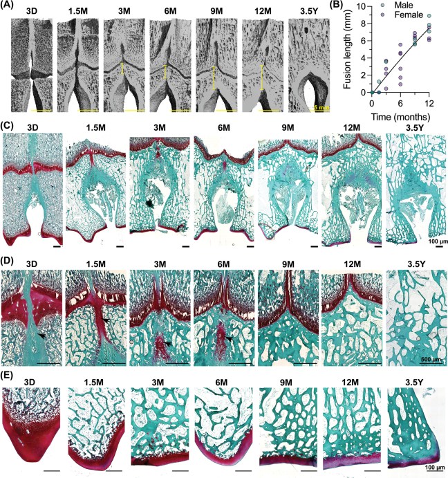
One of the most surprising results related to the synostosis (fusion) of the ends of the distal metacarpus. Unlike humans, the goat forelimb has two metacarpal rudiments (early bones) during embryonic development, and these rudiments undergo synostosis prenatally to form one metacarpal bone at birth. It is thought that the fusion of these bones brings about greater strength in bones especially for activities involving a lot of bending. Fusion happens both in the straight part of the bone (the diaphysis) and also at the joint end (the epiphysis). Fusion in the diaphysis happens prenatally, but we found that – for the first time- that fusion of the ends of the bone happened postnatally, prior to skeletal maturity, through an interesting type of bone formation.
