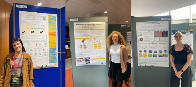
We were delighted to see our very own Giulia, Jemma, and Karin represent the Developmental Biomechanics Group at this year’s Conway Festival of Research and Innovation at UCD. The two-day event was packed with engaging talks, inspiring posters, and plenty of good, solid science. The whole group was in attendance and made the most of the opportunity to connect with other researchers across Conway.
Giulia presented her poster, “A proteomic roadmap of extracellular matrix maturation in developing articular cartilage.” Her work identified around 800 proteins across seven postnatal stages of goat skeletal maturation, revealing a fascinating shift in cartilage composition over time. Early-stage cartilage was enriched with transient collagens and glycoproteins, while later stages showed increased regulators and stabilising proteins; marking a clear transition from matrix assembly to long-term tissue maintenance.
Jemma’s poster, “Mechanisms Underlying the Mechanoregulation of Immature Skeletal Tissues,” showcased her recent progress in developing and validating a single-cell RNA sequencing protocol to study how differential mechanical loading affects key skeletal tissues including the physis, articular cartilage, and ossification centres. Her pilot study provided important validation of the protocol’s robustness and generated valuable preliminary data, and her poster was noted for its clarity and technical depth.
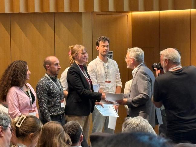
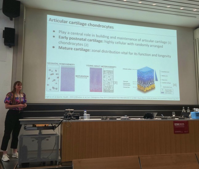
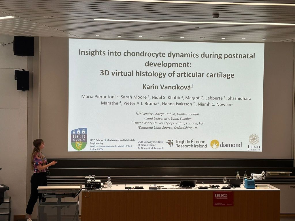
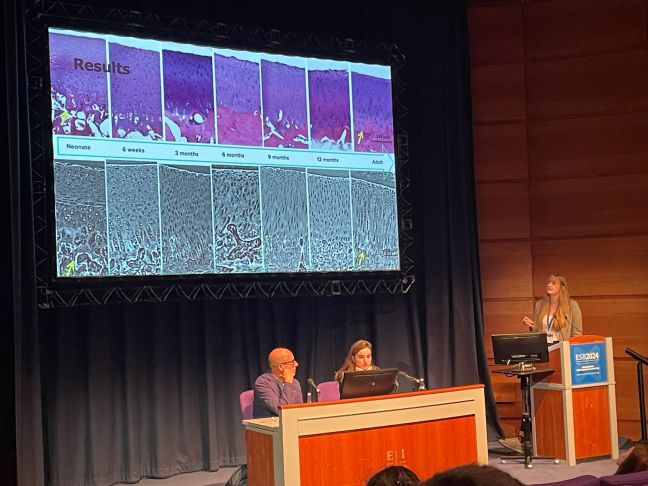
Karin presented “Insights into chondrocyte dynamics during postnatal development: 3D virtual histology of articular cartilage.” Using synchrotron-based phase contrast tomography, she explored developmental changes in chondrocyte organisation and morphology during cartilage maturation; key processes in extracellular matrix formation. Her poster featured striking 3D renderings and heat maps of cell number, volume, and orientation, offering fresh insights into cartilage development in three dimensions. Karin also presented her work as a presentation as part of the PhD/Post-doc seminar series in the school of Mechanical Engineering at UCD.
We’re incredibly proud of Giulia, Jemma, and Karin for showcasing their exciting work and representing the group so brilliantly at this year’s festival.