
Vivien’s final paper from her PhD was published in eCM (open access link). What we found was that when embryos develop without any skeletal muscle, surprisingly, the effects on the skeleton are less severe over development.
Work by our group and others have shown that when skeletal muscle is absent, bones and joints are abnormal with missing cavitation, abnormally shaped bones, and decreased mineralisation. BUT how these abnormalities progress over time in utero was unknown. We wanted to look at stages not previously characterised in detail, and chose TS24 (around e15.5) and TS27 (around e18.5). Our previous work showed different bones and joints are differentially affected by the lack of muscle, so we also looked at a range of rudiments.
At TS24, we found similar effects of absent muscle to those reported before; abnormal sizes and shapes of all major joints in the limb. BUT, at TS27, the joint shapes were much more normal, with much of the significant differences eliminated.

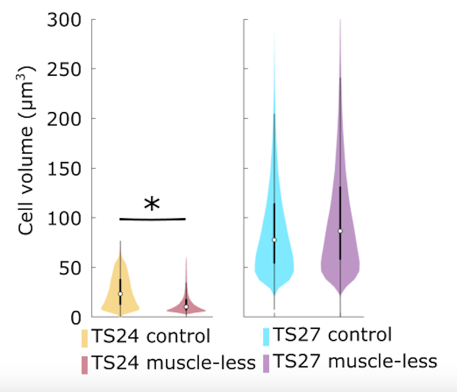
We looked at cell-level activities in the joints at TS24 and TS27 and found that cell size is a possible mechanism underlying the recovery in joint shape over gestation.
Interestingly, cavitation did not change between the two stages, so when cavitation was abnormal at TS24, it didn’t improve by TS27. Therefore, improvements in shape occurred *despite* absent cavitation in some joints.

Perhaps most surprising was the finding that mineralisation, as all muscleless long bones had significantly less mineralisation at TS24, which was completely recovered by TS27, and even exceeded in some rudiments like the ulna. The big question is how does this recovery in both cartilage growth/shape and mineralisation occur? It’s one we are still answering, so watch the space for follow up!
