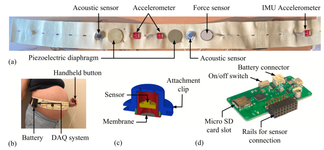
A paper entitled “Multi-modal detection of fetal movements using a wearable monitor” led by Dr Abhishek Ghosh (a former PhD student at Imperial College London, jointly supervised by Prof Nowlan) has been published in Information Fusion.
In the paper, we describe how a prototype wearable fetal movement monitor with multiple different types of vibration sensor was capable of detecting an impressive 82% of maternally sensed fetal movements with an overall accuracy of 90%. The excellent performance of the device was enabled in part by machine learning methods applied to the signals from the different sensors. For more details, please see the open-access paper here.
Congratulations to Abhishek and to all co-authors! Abhishek and Prof Nowlan continue to collaborate through their Wellcome Leap funded project to develop a wearable fetal movement monitor.
Congratulations to Saima whose paper was published in Royal Society Open Science. In the paper, we investigate how mechanical loading from fetal movements affects emergence and maturation of the different types of collagen in the developing skeleton. We found that most collagens were affected by the lack of muscle loading, with collagens II, X and XI being particularly abnormal when skeletal muscle was absent. Using our in vitro bioreactor culture system, we demonstrated that mechanical loading directly modulates the spatial localization and structure of collagens II and X, and excitingly, that mechanical loading in vitro could rescue aspects of the development of collagens II and X from the effects of fetal immobility.
Saima’s imaging is really beautiful- check out the full paper which is available open access here.
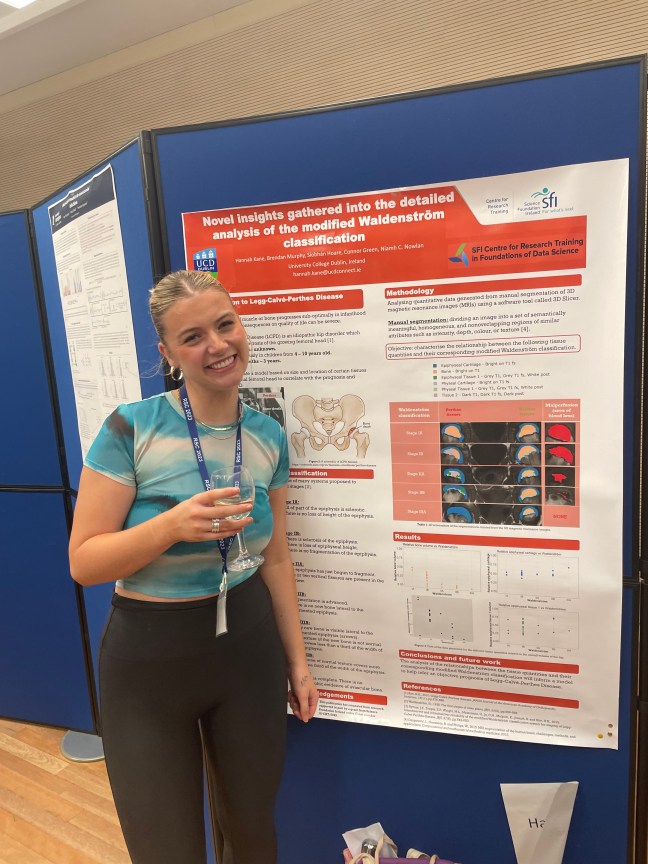
Congratulations to PhD Student Hannah Kane who won “Best Poster” prize at the 46th Research Students’ Conference in Probability and Statistics (RSC 2023) held in Sheffield, UK, on the 11-14th September 2023. Hannah’s poster was entitled “Novel insights gathered into the detailed analysis of the modified Waldenström classification”. Well done Hannah!
Funding worth in excess of €1.25 million from Science Foundation Ireland was announced for the project led by Prof Nowlan and Prof Brama was announced by Minister Simon Harris. The project entitled “Developmentally inspired approaches to cartilage defect healing” will be conducted over five years.
When injury to adult articular cartilage occurs, the defects do not heal, increasing the risk of joint disease and subsequent pain and suffering in humans and animals. In contrast, defects in immature cartilage can heal spontaneously, but there is little understanding of why only immature cartilage heals. Our aim is to recapitulate what happens in immature cartilage to heal adult cartilage defects. We will study which defects heal, and how, depending on animal age. We will use this knowledge to develop novel, ground-breaking treatments for cartilage repair in adult animals and humans.
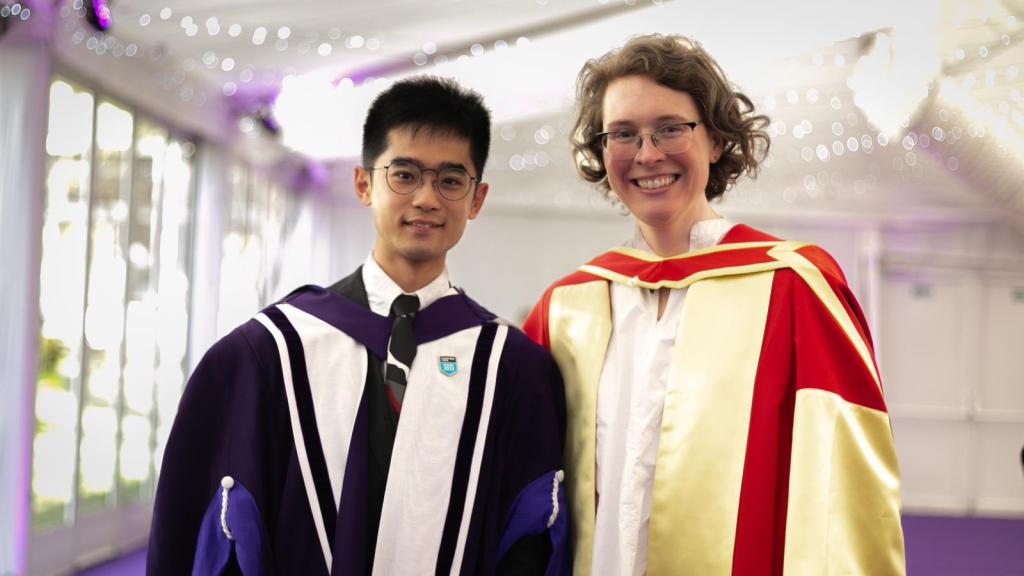
Huge congratulations to Dr Yuming Huang who was conferred with his doctoral degree in the Royal Albert Hall in London on the 3rd May 2023. Niamh was delighted to be there for the lovely ceremony and to celebrate Yuming’s achievements.
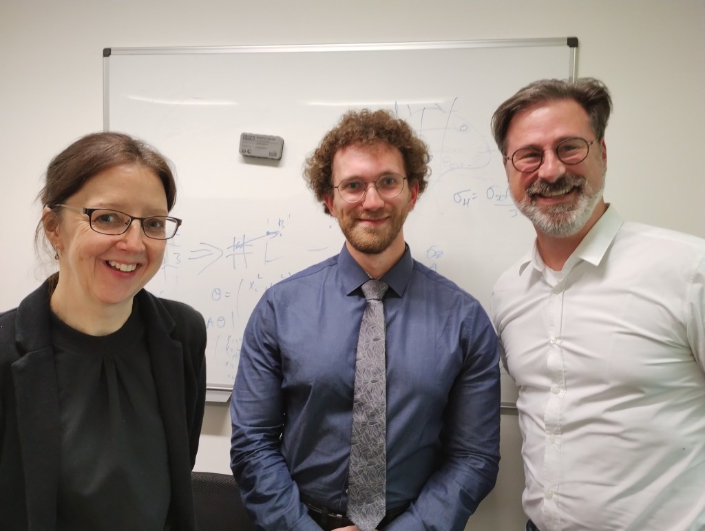
Many congratulations to Jo and James who both passed their PhD vivas with minor corrections! 🤩 Thank you to all the examiners (Dr Kyra Campbell, Dr Choon Hwai Yap, Dr Prof Darryl Overby & Prof Sarah Waters) for their sterling efforts in putting Jo and James through their paces. Thank you too to former group members who rejoined us for the celebrations 🥳
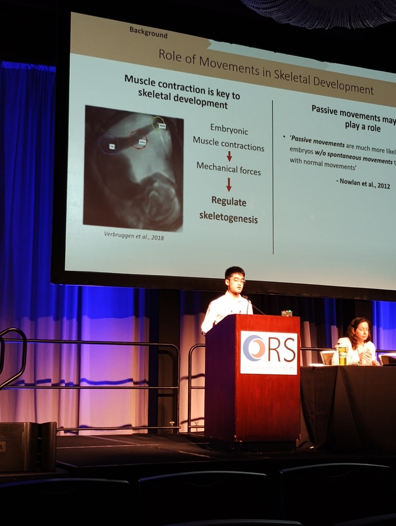
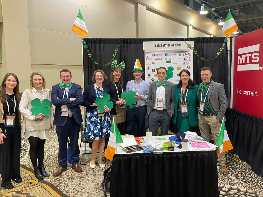
James, Yuming and Niamh, together with alumni Nidal, Rebecca and Stefaan attended the 2023 Annual Meeting of the ORS in Dallas, Texas. James presented a poster, and Yuming and Niamh presented podium talks. Ireland was well represented as the 2023 Guest Nation!
James presenting his posterYuming’s podium presentationSome of the Irish representatives as part of the Guest Nation. Photo credit @LaoiseMcnamara
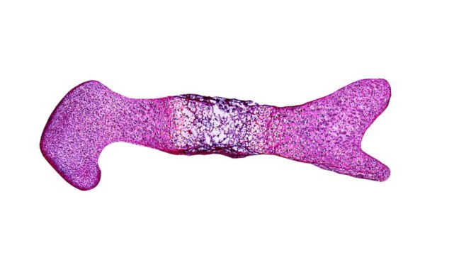
Nidal’s paper entitled “Mechanoregulatory role of TRPV4 in prenatal skeletal development” was published in leading journal Science Advances! The project was a fantastic team effort from Nidal, James, Yuming and Saima, together with collaborator in Trinity College Dublin David Hoey. This work was supported by European Research Council under the European Union’s Seventh Framework Program, ERC Grant agreement number 336306
The movements of a baby in the womb (fetal movements) are a critical sign of the baby’s health and development. Such movements are also important for development of the baby’s bones and joints. When a baby doesn’t move enough, or their movements are restricted in some way, the shapes of their joints don’t form correctly, leading to conditions such as developmental dysplasia of the hip (where the hip joint is unstable or dislocated) or arthrogryposis, where multiple joints are angled abnormally. There is a link between the mechanical forces caused by fetal movements and the processes by which the skeleton takes its shape, but the mechanisms underlying this relationship are unknown.
In this paper, we found that a particular ion channel called TRPV4 (transient receptor potential cation channel subfamily V member 4) is involved in the response of the growing skeleton to the mechanical forces caused by fetal movements. We discovered this link by blocking activity of TRPV4 in embryonic mouse limbs, and showing that the normal response of the tissues to mechanical loading was eliminated. We also showed that activation of TRPV4 by mechanical loading affects proliferation of cells and the production of matrix in the cartilage, both of which affect growth of the joint.
A fascinating thing about TRPV4 is that when the gene which codes for the TRPV4 protein is mutated, a range of different severe skeletal conditions can occur including lethal metatropic dysplasia, spondylometaphyseal dysplasia (dwarfism), and autosomal dominant brachyolmia. Our study is the first to demonstrate that TRPV4 activity in the developing skeleton is closely linked to the mechanical loading from fetal movements. Drugs aimed at targeting TRPV4 are being trialled for a range of different conditions including osteoarthritis and heart failure, and we believe that our research indicates that TRPV4 may be a valuable target for future therapeutic disease modifying drugs for abnormalities of paediatric skeletal development, particularly when fetal movements are reduced or restricted.
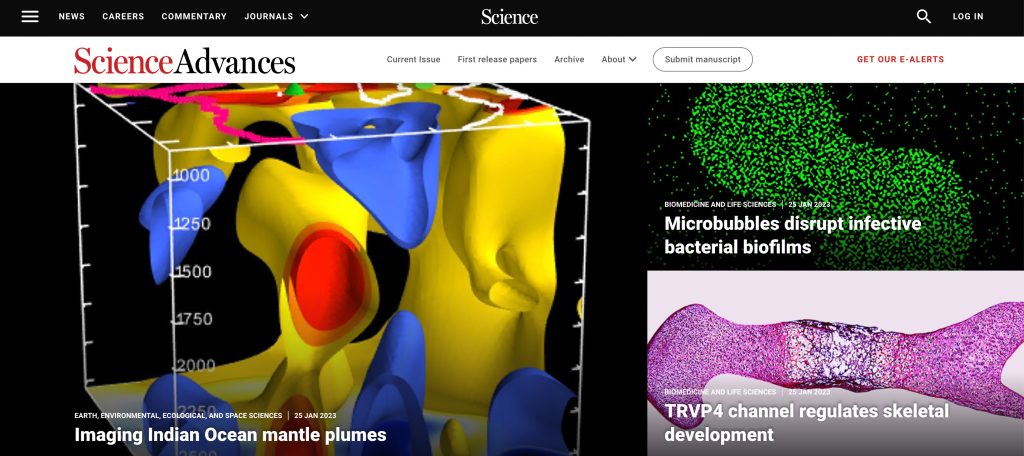
The paper was featured on the Science Advances homepage! Many congratulations to Nidal and all co-authors.
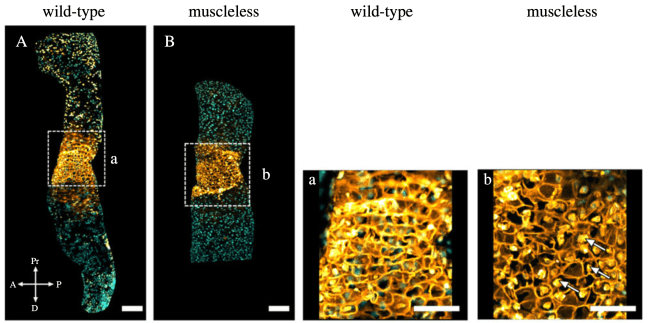
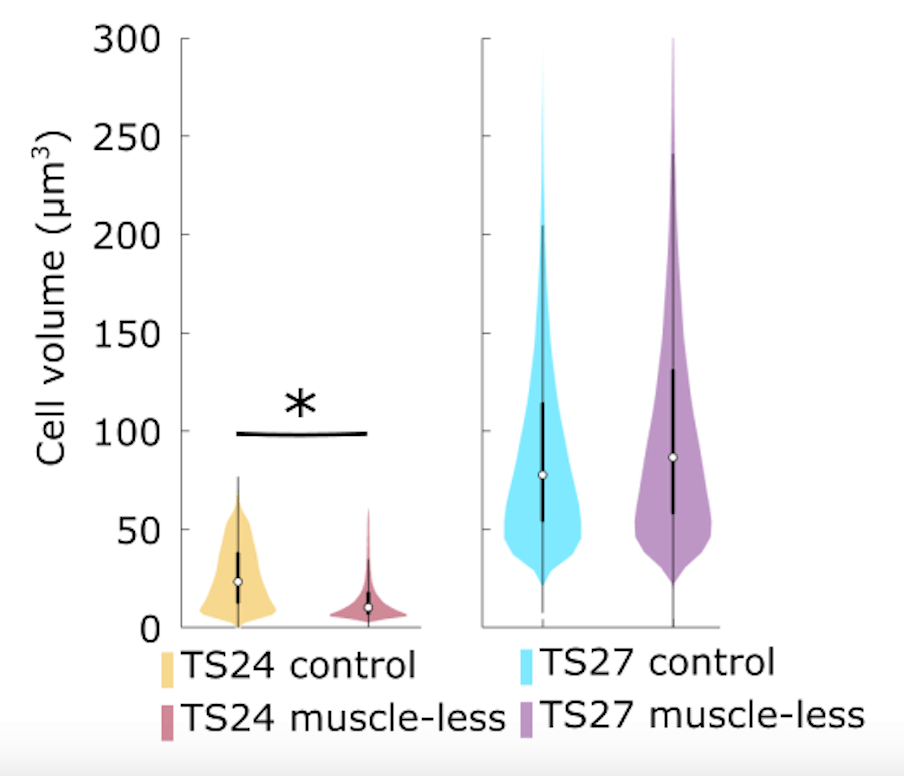
Vivien’s final paper from her PhD was published in eCM (open access link). What we found was that when embryos develop without any skeletal muscle, surprisingly, the effects on the skeleton are less severe over development.
Work by our group and others have shown that when skeletal muscle is absent, bones and joints are abnormal with missing cavitation, abnormally shaped bones, and decreased mineralisation. BUT how these abnormalities progress over time in utero was unknown. We wanted to look at stages not previously characterised in detail, and chose TS24 (around e15.5) and TS27 (around e18.5). Our previous work showed different bones and joints are differentially affected by the lack of muscle, so we also looked at a range of rudiments.
At TS24, we found similar effects of absent muscle to those reported before; abnormal sizes and shapes of all major joints in the limb. BUT, at TS27, the joint shapes were much more normal, with much of the significant differences eliminated.
We looked at cell-level activities in the joints at TS24 and TS27 and found that cell size is a possible mechanism underlying the recovery in joint shape over gestation.
Interestingly, cavitation did not change between the two stages, so when cavitation was abnormal at TS24, it didn’t improve by TS27. Therefore, improvements in shape occurred *despite* absent cavitation in some joints.
Perhaps most surprising was the finding that mineralisation, as all muscleless long bones had significantly less mineralisation at TS24, which was completely recovered by TS27, and even exceeded in some rudiments like the ulna. The big question is how does this recovery in both cartilage growth/shape and mineralisation occur? It’s one we are still answering, so watch the space for follow up!
A baby is stillborn every 16 seconds, leading to heartbreak for more than two million families worldwide per year. Despite advances in care for babies after birth, progress towards reducing the number of stillbirths is lagging behind. Over 50 per cent of stillbirths are associated with a reduction in the baby’s movements in the womb but there is currently no way to track a baby’s movements at home.
Prof Nowlan, together with her collaborators Prof Ravi Vaidyanathan, Prof Christoph Lees & Mr Abhishek Ghosh (Imperial College London) and Prof Fionnuala McAuliffe (UCD) has been awarded a contract as part of Wellcome Leap’s In Utero programme, which aims to create the scalable capacity to measure, model and predict gestational development with a primary goal to reduce stillbirth rates by half. Wellcome Leap is a non-profit organisation founded by the Wellcome Trust to accelerate and increase the number of breakthroughs in human health globally. The team aims to determine how their monitor (called the FM monitor) can be used to measure a baby’s health in the womb. The FM monitor could potentially identify babies who are at risk of stillbirth and will also offer reassurance when the baby is healthy, thereby decreasing the rates of unnecessary induction of labour and early delivery.